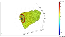
Finite Element Simulation of Shear Wave Propagation Induced by a VCTE Probe
The Fibroscan® (Echosens, Paris, France) device based on vibration–controlled transient elastography (VCTE) is used to non– invasively assess liver stiffness correlated to the hepatic fibrosis. Stiffness is quantified by measuring the velocity of a low–frequency shear wave traveling through the liver, which is proportional to the Young’s modulus E. It has been demonstrated that E is highly correlated with liver fibrosis stage as assessed by liver biopsy. To study the emergence of a two shear wave with different velocity in liver detected with the Fibroscan® on different in vivo cases, simulations with finite element models (FEM) on a 3D anatomical model of liver and ribs can help to understand this propagation patterns. Indeed, the shape and the direction of the shear wave front induced by the Fibroscan® probe in the liver are not entirely known.

Download
- audiere_presentation.pdf - 1.98MB
- audiere_paper.pdf - 0.16MB
