

A key challenge in medical device design is mitigating the risk of blood damage. Modeling and simulation enables engineers to estimate the likelihood of blood damage, improving device safety and performance while reducing computational costs. Veryst Engineering, a COMSOL Certified Consultant, used the COMSOL Multiphysics® software to build surrogate models for predicting recirculation zones and blood damage — specifically, hemolysis — based on a benchmark nozzle geometry from the U.S. Food and Drug Administration (FDA) (Ref. 1).
Matthew Hancock, PhD, principal at Veryst Engineering, said, “As AI and machine learning become part of the regulatory submission process, the real test isn’t just how accurate a surrogate model is compared to the reference simulation; it’s whether we can show, not just assume, that any AI-introduced error is small enough to preserve the integrity of the decision it’s informing. We call this decision reliability.”
Using Simulation to Mitigate Risk
When medical devices interact with blood, there is a risk of them causing unintentional blood damage. This risk poses a design challenge for devices such as instrumentation loops or device–blood vessel connections, including pacemakers, heart valves, and catheters.
Hemolysis occurs when red blood cells rupture and release hemoglobin into the surrounding blood plasma. When modeling medical devices, this type of blood damage can be measured by the increase in unbound hemoglobin in the blood circulating through the device. Quantifying the hemoglobin, while considering sublethal damage and thrombosis, can lead to more informed design decisions.
Veryst’s goal was to create a simulation that can aid medical device designers in understanding the relationship between device geometry and blood damage. To meet this goal, the team turned to multiphysics simulation and surrogate models, which also offer the benefit of significantly reducing the resources needed to conduct real-world experiments.
In a poster presentation from a COMSOL Conference, engineers at Veryst shared how they trained and deployed surrogate models to provide visualizations of blood damage in real time. They were able to change aspects of the design and different operating parameters and observe the effects that occurred. These analyses helped them determine the most influential parameters and their tolerance levels.
Building and Training Surrogate Models
For their simulation work, the Veryst engineers used deep neural network (DNN) surrogate models to predict blood velocity and hemolysis. This work references and builds upon Veryst’s previous work modeling blood damage in the FDA benchmark nozzle (Ref. 2), which was presented in an invited talk at the COMSOL Conference 2020. The team was able to use that data from the previous full-model simulations to help train the DNN models.
Additionally, the FDA has benchmark datasets for validating numerical simulations of blood flow through medical devices (Ref. 3), and requirements must be met for a medical device to receive FDA approval. One benchmark dataset is for a nozzle that has a tube with a contraction, neck, and expansion, or a conical change in diameter at one end of the throat and sudden change at the other end (Figure 1).
In order to prepare their nozzle model for submission to the FDA, the Veryst engineers chose fluid properties that match the FDA benchmark protocol. They set up four adjustable input parameters:
- Flow rate
- Converging length
- Neck diameter
- Diverging length
The surrogate model predicts the blood velocity and estimates hemolysis using a power-law relationship as a function of stress (Ref. 2). The hemolysis model output represents the prediction of how much hemoglobin is released to the blood stream due to cell rupture or leakage.
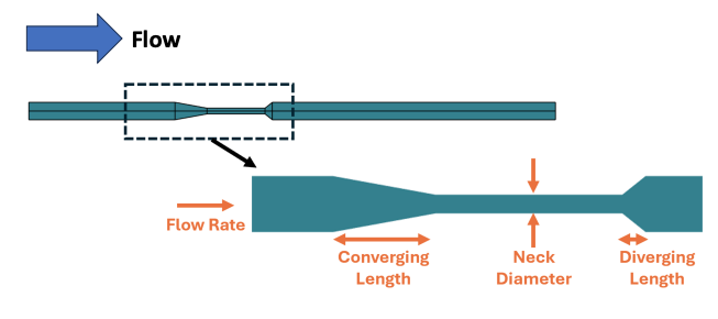 Figure 1. A diagram showing nozzle setup and inputs for the DNN model.
To build the hemolysis model, the team computed mean hemolysis across the flow’s exit with a design of experiments sample of 9000 simulations. A surrogate model was built to train the velocity components u and w on raw data. The DNN surrogate model included 3 hidden layers, each with 20 nodes using tanh activation functions. To train the model, 5000 epochs were used. The velocity network included a DNN trained on 500 simulations at 3500 epochs, with 32 nodes in each of the 3 hidden layers. The velocity training data utilized all mesh points after expansion, with the diverging length set to 0 in order to focus on recirculation zones.
Predictions Align with Previous Data
After creating a surrogate model to train u and w, the team developed a modified DNN with a physically informed no-slip hard constraint. This network was trained on the deviation of axial velocity, w’, from the analytic laminar flow solution w’ = w – (2*Q/A)(1–(r/R)2). In this DNN, Q represents the flow rate, A represents the cross-sectional area, r represents the radial coordinate, and R represents the end radius (Figure 2).
The raw data DNN and the hard-constrained DNN both accurately replicate flow profiles in turbulent circumstances, which represent most of the underlying training data. These DNNs are less quantitative when it comes to matching the laminar simulation or the transition to turbulence regimes.
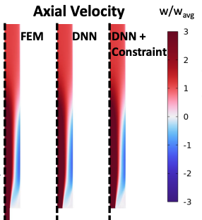
Figure 2. Velocity at Reynolds number Re = 6500, which replicates the FDA geometry, showing recirculation after expansion.
When the hemolysis predictions are trained to match the log of hemolysis, the results are within 10% accuracy across a wide range of inputs. Veryst found that changing the converging and diverging lengths of the geometry had minimal effect on hemolysis in both the finite element analysis (FEA) and DNN predictions. To validate the surrogate model, the team compared the hemoglobin concentrations predicted by FEA and the DNN for parameters based on the experiments of Herbertson (Ref. 4)(Figure 3).
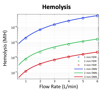
Figure 3. The fraction of released hemoglobin at the nozzle endpoint for the FDA geometry with variable neck radius and flow rate.
“Getting hemolysis predictions within 10% of the full CFD model, while cutting runtime from hours to milliseconds, is what makes real-time design exploration possible,” said Joseph Barakat, PhD, lead engineer at Veryst Engineering.
Veryst’s work is based around the publicly available FDA benchmark geometry, but it could be expanded for a wide variety of future uses. The methodology could potentially be used to estimate blood damage in a range of medical devices, such as instrumentation loops and device–blood vessel connections.
Hancock added, “Formally combining surrogate approximation error, numerical error, and validation discrepancy into a single total-error estimate to assess this decision reliability — grounded in ASME’s Verification and Validation 20 and 40 standards and the broader verification, validation, and uncertainty quantification literature — is a natural next step, particularly as the FDA’s Center for Devices and Radiological Health continues building out regulatory science tools for AI-accelerated device simulation.”
Next Step
Want to learn more about Veryst’s work on predicting blood damage with surrogate models? Click the button below to see the team’s poster from the COMSOL Conference:
References
- A. Spann, J. Barakat, and M. Hancock, “Deep Neural Network Surrogate Model for Blood Damage Modeling in FDA Hemolysis Benchmark,” COMSOL Conference 2024 Boston, 2024; https://www.comsol.com/paper/deep-neural-network-surrogate-model-for-blood-damage-modeling-in-fda-hemolysis-benchmark-136422
- A. Kermani, A. Vanegas, and A. Spann, “Blood Damage Modeling of FDA Benchmark Nozzle,” COMSOL Conference 2020 Boston, 2020; https://www.comsol.com/paper/blood-damage-modeling-of-fda-benchmark-nozzle-93171
- U.S. Food and Drug Administration, Benchmark dataset for validating computational fluid dynamic (CFD) simulation of blood flow through generalized medical device geometries (RST24CV11.01), 2024; https://cdrh-rst.fda.gov/benchmark-dataset-validating-computational-fluid-dynamic-cfd-simulation-blood-flow-through
- L.H. Herbertson et al., “Multilaboratory study of flow-induced hemolysis using the FDA benchmark nozzle model,” Artificial Organs, vol. 39, no. 3, pp. 237–248, 2015; https://pubmed.ncbi.nlm.nih.gov/25180887/





Comments (0)